VIVO Pathophysiology
The Gastrointestinal Immune System
The lumen of the gastrointestinal tract is outside of the body and much of it is heavily populated with potentially pathogenic microorganisms. It is thus important that the immune system establish and maintain a strong presence at this mucosal boundary, and indeed, the digestive tube is heavily laden with lymphocytes, macrophages and other cells that participate in immune responses.
Aside from all of its other functions, the gastrointestinal tract is a lymphoid organ, and the lymphoid tissue within it is collectively referred to as the gut-associated lymphoid tissue or GALT. The number of lymphocytes in the GALT is roughly equivalent to those in the spleen, and, based on location, these cells are distributed in three basic populations:
Peyer's Patches: These are lymphoid follicles similar in many ways to lymph nodes, located in the mucosa and extending into the submucosa of the small intestine, especially the ileum. In adults, B lymphocytes predominate in Peyer's patches. Smaller lymphoid nodules can be found throughout the intestinal tract.
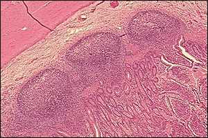
In the image of canine ileum below, three lymphoid follicles of a Peyer's patch can be seen. The muscularis is at the top left, and mucosal epithelium in the bottom right.
Lamina propria lymphocytes: These are lymphocytes scattered in the lamina propria of the mucosa. A majority of these cells are IgA-secreting B cells.
Intraepithelial lymphocytes: These are lymphocytes that are positioned in the basolateral spaces between lumenal epithelial cells, beneath the tight junctions (they are "inside" the epithelium, but not inside epithelial cells as the name may incorrectly suggest).
Another important component of the GI immune system is the M or microfold cell. M cells are a specific cell type in the intestinal epithelium over lymphoid follicles that endocytose a variety of protein and peptide antigens. Instead of digesting these proteins, M cells transport them into the underlying tissue, where they are taken up by local dendritic cells and macrophages.
Dendritic cells and macrophages that receive antigens from M cells present them to T cells in the GALT, leading ultimately to appearance of immunoglobulin A-secreting plasma cells in the mucosa. Dendritic cells below the epithelium can also sample lumenal antigens by pushing pseudopods between epithelial cells. The secretory IgA is transported through the epithelial cells into the lumen, where, for example, it interferes with adhesion and invasion of bacteria.
T cells exposed to antigen in Peyer's patches also migrate into the lamina propria and the epithelium, where they mature to cytotoxic T cells, providing another mechanism for containing microbial assaults.
In addition to the GALT discussed above, lymph nodes that receive lymph draining from the gut (mesenteric nodes) and Kupffer cells (phagocytic cells in the liver) play important roles in protecting the body against invasion.
References and Reviews
- Nagler-Anderson C: Man the barrier! Strategic defences in the intestinal mucosa. Nat Rev Immunol 1:59-67, 2001.
- Neutra MR, Frey A, Kraehenbuhl JP: Epithelial M cells: Gateways for mucosal infection and immunization. Cell 86:345, 1996.
Send comments to Richard.Bowen@colostate.edu