VIVO Pathophysiology
Physiology of the Hepatic Vascular System
Hepatic Blood Volume and Reservoir Function
The liver receives approximately 30% of resting cardiac output and is therefore a very vascular organ. The hepatic vascular system is dynamic, meaning that it has considerable ability to both store and release blood - it functions as a reservoir within the general circulation.
In the normal situation, 10-15% of the total blood volume is in the liver, with roughly 60% of that in the sinusoids. When blood is lost, the liver dynamically adjusts its blood volume and can eject enough blood to compensate for a moderate amount of hemorrhage. Conversely, when vascular volume is acutely increased, as when fluids are rapidly infused, the hepatic blood volume expands, providing a buffer against acute increases in systemic blood volume.
Formation of Lymph in the Liver
Approximately half of the lymph formed in the body is formed in the liver. Due to the large pores or fenestrations in sinusoidal endothelial cells, fluid and proteins in blood flow freely into the space between the endothelium and hepatocytes (the "space of Disse"), forming lymph. Lymph flows through the space of Disse to collect in small lymphatic capillaries associated with portal triads (the reason they are not called portal tetrads is because these lymphatic vessels are virtually impossible to identify in standard histologic sections), and from there in the systemic lymphatic system.
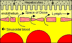
As you might expect, if pressure in the sinusoids increases much above normal, there is a corresponding increase in the rate of lymph production. In severe cases the liver literally sweats lymph, which accumulates in the abdominal cavity as ascitic fluid. What lesions can you envision that would raise blood pressure in sinusoids, resulting in production of ascites ?
The Hepatic Phagocytic System
The liver is host to a very important part of the phagocytic system. Lurking in the sinusoids are large numbers of a type of tissue macrophage known as the Kupffer cell. Kupffer cells are actively phagocytic and represent the main cellular system for removal of particulate materials and microbes from the circulation. The image below is a lightly stained section of liver from a mouse that was injected intravenously with a very small quantity of India ink - Kupffer cells are clearly visible throughout the section because they have phagocytosed the ink particles and appear dark black.
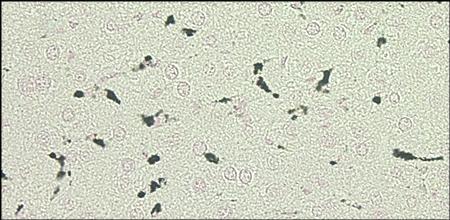
Their location just downstream from the portal vein allows Kupffer cells to efficiently scavenge bacteria that get into portal venous blood through breaks in the intestinal epithelium, thus preventing invasion of the systemic circulation.
Send comments to Richard.Bowen@colostate.edu