
| Reproduction Index | Glossary |
|---|
|
Placental Structure and Classification |
The placentas of all eutherian (placental) mammals provide common structural and functional features, but there are striking differences among species in gross and microscopic structure of the placenta. Two characteristics are particularly divergent and form bases for classification of placental types:
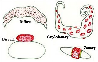
Differences in these two properties allow classification of placentas into several fundamental types. Classification Based on Placental Shape and Contact PointsExamination of placentae from different species reveals striking differences in their shape and the area of contact between fetal and maternal tissue:
 Classification Based on Layers Between Fetal and Maternal BloodJust prior to formation of the placenta, there are a total of six layers of tissue separating maternal and fetal blood. There are three layers of fetal extraembryonic membranes in the chorioallantoic placenta of all mammals, all of which are components of the mature placenta:
There are also three layers on the maternal side, but the number of these layers which are retained - that is, not destroyed in the process of placentation - varies greatly among species. The three potential maternal layers in a placenta are:
 One classification scheme for placentas is based on which maternal layers are retained in the placenta, which of course is the same as stating which maternal tissue is in contact with chorionic epithelium of the fetus. Each of the possibilities is observed in some group of mammals. |
| Type of Placenta | Maternal Layers Retained | Examples | ||
|---|---|---|---|---|
| Endometrial Epithelium |
Connective Tissue |
Uterine Endothelium |
||
| Epitheliochorial | + | + | + | Horses, swine, ruminants |
| Endotheliochorial | - | - | + | Dogs, cats |
| Hemochorial | - | - | - | Humans, rodents |
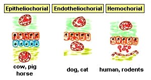 In humans, fetal chorionic epithelium is bathed in maternal blood because chorionic villi have eroded through maternal endothelium. In contrast, the chorionic epithelium of horse and pig fetuses remains separated from maternal blood by 3 layers of tissue. One might thus be tempted to consider that exchange across the equine placenta is much less efficient that across the human placenta. In a sense this is true, but other features of placental structure make up for the extra layers in the diffusion barrier; it has been well stated that "The newborn foal provides a strong testimonial to the efficiency of the epitheliochorial placenta." Summary of Species Differences in Placental ArchitectureThe placental mammals have evolved a variety of placental types which can be broadly classified using the nomenclature described above. Not all combinations of those classification schemes are seen or are likely to ever be seen - for instance, no mammal is known to have a diffuse, endotheliochorial, or a hemoendothelial placenta. Placental types for "familiar" mammals are summarized below, with supplemental information provided for a variety of "non-familiar" species. |
| Type of Placenta | Common Examples |
|---|---|
| Diffuse, epitheliochorial | Horses and pigs |
| Cotyledonary, epitheliochorial | Ruminants (cattle, sheep, goats, deer) |
| Zonary, endotheliochorial | Carnivores (dog, cat, ferret) |
| Discoid, hemochorial | Humans, apes, monkeys and rodents |
ResourcesComparative Placentation is an excellent and comprehensive site for obtaining information on placental structure for a large number of domestic and wild animals |
| Index of: Implantation and Development of the Placenta | |||
 |
Attachment and Implantation | Transport Across the Placenta |  |
Last updated on September 25, 2011 |
| Author: R. Bowen |
| Send comments via form or email to rbowen@colostate.edu |