
| Reproduction Index | Glossary |
|---|
|
Endometrial Cups and Secretion of Equine Chorionic Gonadotropin |
Shortly after establishment of pregnancy in equids, high concentrations of the hormone equine chorionic gonadotropin (eCG) appear in the mare's serum. This hormone is also called pregnant mare's serum gonadotropin and is actually equine luteinizing hormone. The source of eCG is a placenta-associated structure called an endometrial cup, which is derived from the fetus, forms several weeks into gestation, and is immunologically destroyed 2 to 3 months later. 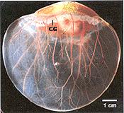
Endometrial cups develop from cells of the chorionic girdle, which can first be detected histologically at roughly 25 days of gestation. Initially, this structure is a narrow band of thicked trophoblast that develops circumferentially around the conceptus at a point where the membranes of the allantois and yolk sac meet. Trophoblastic epithelial cells of the girdle proliferate to form ridges, and later glands, that abut against and are flattened by the surface of the endometrium. A mucoid material secreted into the glands adheres to the endometrium and tends to hold the girdle in place. In the image to the right, the chorionic girdle (CG) can be seen encircling the upper portion of the embryo (kindly provided by Dr. O. J. Ginther). Beginning on days 36 to 38 of gestation, hyperplastic girdle cells begin to rapidly invade and destroy underlying endometrium. In this process they denude surface endometrial epithelial cells and migrate down into endometrial glands, eventually breaking through the basement membrane and invading into the underlying stroma. Within 2 to 3 days after entering the uterine stroma, they round up and differentiate into mature eCG-secreting endometrial cup cells. Girdle cell invasion and proliferation result in formation of tightly packed mass of trophoblast-derived cells containing little stroma - these are the endometrial cups. Invasion of endometrial glands leads to destruction of their apical epithelium; deeper segments of those glands are spared, but their lumens are obstructed by cup cells and they become distended with secretions. Endometrial cups reach their maximum size and eCG output about 55 to 70 days into gestation, at which time they appear as pale, circular or U-shaped plaques on the surface of the endometrium. Their size and shape varies tremendously, from approximately 1 cm circles to ribbons of tissue at least 10 cm in length. At this mature stage, the differentiated trophoblast cells are distinctive - they are round and almost always binucleate. The images below show endometrial cups (EC) at three stages of gestation. In the rightmost image, umbilical vessels are seen at the bottom. 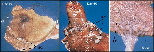 Development of the cups is paralleled almost from their beginning by a striking maternal cellular immune response. This response is initially seen as an accumulation of T lymphocytes at the periphery of the cups, and progresses to a massive accumulation of T cells, B cells, macrophages and other leukocytes in the stroma surrounding the cups. After day 70 to 80, these leukocytes begin to invade the destroy the base of the cup. Eventually, the entire cup is destroyed and sloughs completely from the endometrial surface; the time of this event varies significantly among mares, but usually occurs between days 100 and 140. Immunological destruction of the endometrial cups appears to be a response to paternal class I MHC antigens, which are highly expressed on invading girdles cells. In conjunction with the cellular response is a vigorous humoral immune response to these antigens. Several interesting observations observations on endometrial cup biology have been made in interspecific equine pregnancies. In mares carrying donkey conceptuses, the chorionic girdle fails to invade the endometrium, and endometrial cups do not develop. Most of these pregnancies are aborted between days 80 and 90, but the roughly 30% that survive and are carried to term do so in the absense of eCG. However, in donkeys carrying a hinney fetus, the cups develop to a much larger size and considerably higher concentrations of eCG are achieved than in donkeys carrying a donkey fetus.
|
| Index of: Implantation and Development of the Placenta |
Last updated on March 14, 2000 |
| Author: R. Bowen |
| Send comments via form or email to rbowen@colostate.edu |
 References and Reviews