VIVO Pathophysiology
Histology of the Adrenal Medulla
The most abundant cell in the adrenal medulla is the chromaffin cell. That name derives from the phenomenon, observed long ago, that if adrenal gland is fixed in a solution containing chromium salts, it takes on a brownish appearance due to oxidation of catecholamines to melanin. Chromaffin cells are also referred to by some as pheochromocytes.
Chromaffin cells are columnar in shape and rather basophilic. At higher magnification, they are seen to have a granular cytoplasm due to hormone-containing granules. They are arranged in clusters, usually around medullary veins, as seen below in an image of rabbit adrenal (H&E stain).
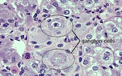
The adrenal medulla is richly innervated by preganglionic sympathetic fibers. Additionally, small numbers of sympathetic ganglion cells are commonly observed in the medulla. Ganglion cells are round or polygonal with prominent nuclei. A cluster of ganglion cells is seen in the image below.
Send comments to Richard.Bowen@colostate.edu