VIVO Pathophysiology
Histology of the Adrenal Cortex
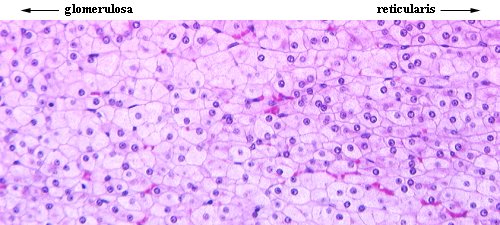
Cells in the adrenal cortex are arranged into three concentric zones. At both low and high magnification, one can readily differentiate these zones based on the pattern produced by cords of cells. However, the boundaries between zones are indistinct.
The outermost zone is the zona glomerulosa. Cells within this zone tend to be columnar in shape and are arranged in irregular cords. In some species, cells adjacent to the capsule are are arranged in quite regular "arcades". In the image below, the zona glomerulosa from the adrenal of a cat and rabbit is shown (H&E stain).

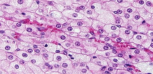
The zona fasiculata is the middle and largest of the three zones in the cortex. Cells in the fasiculata are polyhedral and usually have a foamy appearance due to abundant lipid droplets. They also are arranged in distinctively straight cords that radiate toward the medulla. The image below is of the fasiculata in a rabbit adrenal (H&E stain).
As seen the next image, cortical capillaries are usually prominent within the fasiculata. Notice the shape and foamy appearance of the cells in this micrograph.
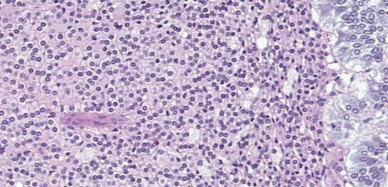
The innermost zone of the cortex is the zona reticularis. Cells within this zone are arranged in cords that project in many different directions and anastomose with one another. In the image below, also of a rabbit adrenal, the reticularis occupies roughly 2/3rds of the center, with a bit of fasiculata on the left and the medulla on the right (H&E).
Send comments to Richard.Bowen@colostate.edu