VIVO Pathophysiology
Gross and Microscopic Anatomy of the Stomach
|
The stomach is an expanded section of the digestive tube between the esophagus and small intestine. It's characteristic shape is shown, along with terms used to describe the major regions of the stomach. The right side of the stomach shown above is called the greater curvature and that on the left the lesser curvature. The most distal and narrow section of the stomach is termed the pylorus - as food is liquefied in the stomach it passes through the pyloric canal into the small intestine. The wall of the stomach is structurally similar to other parts of the digestive tube, with the exception that the stomach has an extra, oblique layer of smooth muscle inside the circular layer, which aids in performance of complex grinding motions. | 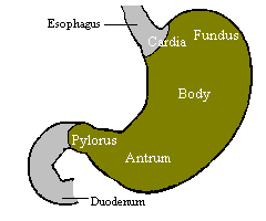 |
|
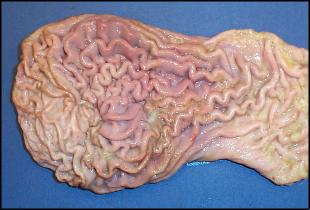
In the empty state, the stomach is contracted and its mucosa and submucosa are thrown up into distinct folds called rugae; when distended with food, the rugae are "ironed out" and flat. The image to the right shows rugae on the surface of a dog's stomach. |
|
|
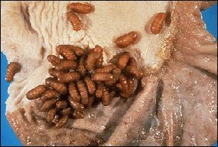
Within the stomach there is an abrupt transition from stratified squamous epithelium extending from the esophagus to a columnar epithelium dedicated to secretion. In most species, this transition is very close to the esophageal orifice, but in some, particular horses and rodents, stratified squamous cells line much of the fundus and part of the body. The image to the right is of the mucosal surface of an equine stomach showing esophageal epithelium (top) and glandular epithelium (bottom). The creatures attached to the surface are bots, larval forms of Gasterophilus. |
|
|
If the lining of the stomach is examined with a hand lens, one can see that it is covered with numerous small holes. These are the openings of gastric pits which extend into the mucosa as straight and branched tubules, forming gastric glands. |
|
Four major types of secretory epithelial cells cover the surface of the stomach and extend down into gastric pits and glands:
There are differences in the distribution of these cell types among regions of the stomach - for example, parietal cells are abundant in the glands of the body, but virtually absent in pyloric glands. The micrograph to the right shows a gastric pit invaginating into the mucosa (fundic region of a raccoon stomach). Notice that all the surface cells and the cells in the neck of the pit are foamy in appearance - these are the mucous cells. The other cell types are farther down in the pit and, in this image, difficult to distinguish. | 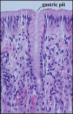 |
Here's an interesting piece of trivia about the stomach: the platypus does not have one. In this strange mammal, the distal esophagus is dilated, but the platypus does not have a glandular stomach. Moreover, its genome has deletions of some of the key genes involved in gastric sections, including those for pepsinogens, the gastric H+/K+ ATPase (proton pump) and the hormone gastrin.
Send comments to Richard.Bowen@colostate.edu