VIVO Pathophysiology
Pancreatic Histology: Endocrine Tissue
If you examine a section of pancreas at low magnification, you will undoubtedly observe distinctive areas of pale staining embedded within the exocrine tissue of lobules (marked with a red * below). These are the Islets of Langerhans, the endocrine component of the pancreas.
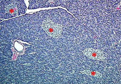
Islets contain several different endocrine cell types. The most abundant are beta cells, which produce insulin, and alpha cells, which secrete glucagon. In sections stained with H&E, the different endocrine cell types cannot be differentiated from one another. Special stains, or better yet, immunostaining, is required to identify specific cell types.
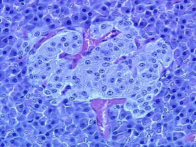
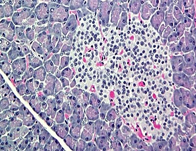
The endocrine cells within islets are arranged as irregular cords around abundant capillaries, which receive the secreted hormones for delivery into the systemic circulation. Examine the images of islets below and note their rich vascularity, evidenced by clusters and streams of red blood cells next to endothelial cells that can be identified by their flattened nuclei.
Equine pancreas (H&E stain)
Monkey pancreas (H&E stain)
Send comments to Richard.Bowen@colostate.edu