VIVO Pathophysiology
Hepatic Histology: Hepatocytes
Hepatocytes are the chief functional cells of the liver and perform an astonishing number of metabolic, endocrine and secretory functions. Roughly 80% of the mass of the liver is contributed by hepatocytes.
In three dimensions, hepatocytes are arranged in plates that anastomose with one another. The cells are polygonal in shape and their sides can be in contact either with sinusoids (sinusoidal face) or neighboring hepatocytes (lateral faces). A portion of the lateral faces of hepatocytes is modified to form bile canaliculi. Microvilli are present abundantly on the sinusoidal face and project sparsely into bile canaliculi.
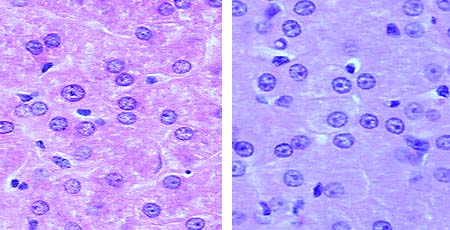
Hepatocyte nuclei are distinctly round, with one or two prominent nucleoli. A majority of cells have a single nucleus, but binucleate cells are common. The micrographs below (H&E stain) demonstrate these features in sections of liver from a pig (left) and raccoon (right).
Hepatocytes are exceptionally active in synthesis of protein and lipids for export. As a consequence of these activities, ultrastructural examination of hepatocytes reveals bountiful quantities of both rough and smooth endoplasmic reticulum. In contrast to most glandular epithelial cells that contain a single Golgi organelle, hepatocytes typically contain many stacks of Golgi membranes. Golgi vesicles are particularly numerous in the vicinity of the bile canaliculi, reflecting transport of bile constituents into those channels.
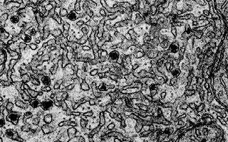
Another important function of hepatocytes is to synthesize and secrete very low density lipoproteins. These complexes are seen in electron micrographs as electron-dense particles within smooth endoplasmic reticulum.
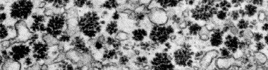
Another type of particle observed in copious quantities in liver is glycogen. Glycogen is a polymer of glucose and the density of glycogen aggregates in hepatocytes varies dramatically depending on whether the liver is examined shortly after a meal (abundant glycogen) or following a prolonged fast (minimal quantities of glycogen). When viewed with an electron microscope, glycogen particles are typically arranged in chrysanthemum-like clusters of electron-dense particles, as seen in the following image:
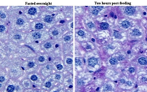
In paraffin sections of liver stained with hematoxylin and eosin, accumulations of glycogen in hepatocytes do not stand out. However, when stained with using the periodic acid-Schiff (PAS) technique, glycogen stains bright pink in color. The images below represent PAS-stained sections of liver from two mice:
- Left panel: from a mouse that fasted overnight and thus had very low levels glycogen in liver.
- Right panel: from mouse that stuffed himself on mouse chow two hours prior to fixing the liver, and thus had high levels of hepatic glycogen. These accumulations are seen as pink areas of PAS-positive material throughout the section.
Back to: Liver Histology: Introduction and Index ^
Send comments to Richard.Bowen@colostate.edu