VIVO Pathophysiology
Hepatic Histology: The Acinus
The hepatic acinus is the functional unit of the liver. The acinus is more difficult to visualize than the lobule, but represents a unit that is of more relevance to hepatic function because it is oriented around the afferent vascular system.
As seen overlayed onto lobules in the diagram below, the acinus consists of an irregular shaped, roughly ellipsoidal mass of hepatocytes aligned around the hepatic arterioles and portal venules just as they anastomose into sinusoids.
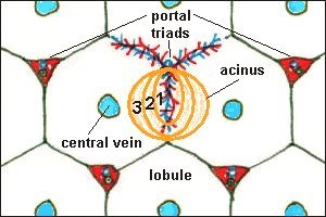
The acinus is roughly divided into zones that correspond to distance from the arterial blood supply - those hepatocytes closest to the arterioles (zone 1 below) are the best oxygenated, while those farthest from the arterioles have the poorest supply of oxygen. This arrangement also means that cells in the center of the acinus (again, zone 1) are the first to "see" and potentially absorb blood-borne toxins absorbed into portal blood from the small intestine.
The net result is that a variety of pathologic processes lead to lesions that reflect acinar structure; for example, necrosis of hepatocytes at the periphery of the acinus.
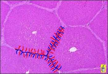
In the image below, a depiction of the arterial and venous blood supply emanating from one triad has been painted onto a micrograph of porcine liver. Try to picture how these vascular branches define acini, then pass your mouse pointer over the image to outline one acinus.
Back to: Liver Histology: Introduction and Index ^
Send comments to Richard.Bowen@colostate.edu