VIVO Pathophysiology
Gross and Microscopic Anatomy of the Large Intestine

The large intestine is that part of the digestive tube between the terminal ileum and anus. Depending on the species, ingesta from the small intestine enters the large intestine through either the ileocecal or ileocolic valve. Within the large intestine, three major segments are recognized:
- the cecum is a blind-ended pouch that in humans carries a worm-like extension called the vermiform appendix.
- the colon constitutes the majority of the length of the large intestine and is subclassified into ascending, transverse and descending segments.
- the rectum is the short, terminal segment of the digestive tube, continuous with the anal canal.
The variation in relative dimension of the large intestine is largely correlated with diet. In herbivores like horses and rabbits which depend largely on microbial fermentation, the large intestine is very large and complex. Omnivores like pigs and humans have a substantial large intestine, but nothing like that seen in herbivores. Finally, carnivores such as dogs and cats have a simple and small large intestine.
There are many similarities in the histologic structure of the mucosa in large and small intestine. The most obvious difference is that the mucosa of the large intestine is devoid of villi. It has numerous crypts which extend deeply and open onto a flat lumenal surface. The stem cells which support rapid and continuous renewal of the epithelium are located either at the bottom or midway down the crypts. These cells divide to populate the cryptal and surface epithelium.
Mucus-secreting goblet cells are also much more abundant in the colonic epithelium than in the small gut.
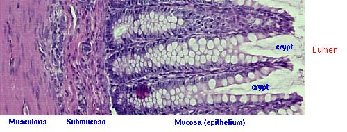
The image above shows a section of colon from a dog. Note the crypts extending from the lumen, and the numerous, foamy goblet cells that populate the epithelium of the crypts.
Send comments to Richard.Bowen@colostate.edu