VIVO Pathophysiology
Digestive Anatomy in Ruminants
The stomach of ruminants has four compartments: the rumen, reticulum, omasum and abomasum, as shown in the following diagram:
Collectively, these organs occupy almost 3/4ths of the abdominal cavity, filling virtually all of the left side and extending significantly into the right. The reticulum lies against the diaphragm and is joined to the rumen by a fold of tissue. The rumen, far and away the largest of the forestomaches, is itself sacculated by muscular pillars into what are called the dorsal, ventral, caudodorsal and caudoventral sacs. In many respects, the reticulum can be considered a "cranioventral sac" of the rumen; for example, ingesta flows freely between these two organs. The reticulum is connected to the spherical omasum by a short tunnel.
The abomasum is the ruminant's true or glandular stomach. Histologically, it is very similar to the stomach of monogastrics.
The interior of the rumen, reticulum and omasum is covered exclusively with stratified squamous epithelium similar to what is observed in the esophagus. Each of these organs has a very distinctive mucosa structure, although within each organ, some regional variation in morphology is observed. The images below are from a sheep.
| The interior surface of the rumen forms numerous papillae that vary in shape and size from short and pointed to long and foliate. | 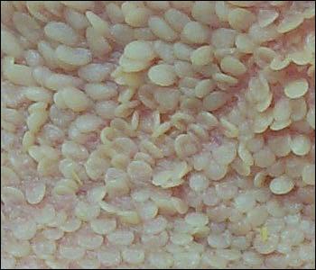 |
| Reticular epithelium is thrown into folds that form polygonal cells that give it a reticular, honey-combed appearance. Numerous small papillae stud the interior floors of these cells. | 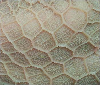 |
| The inside of the omasum is thrown into broad longitudinal folds or leaves reminiscent of the pages in a book (a lay term for the omasum is the 'book'). The omasal folds, which in life are packed with finely ground ingesta, have been estimated to represent roughly one-third of the total surface area of the forestomachs. | 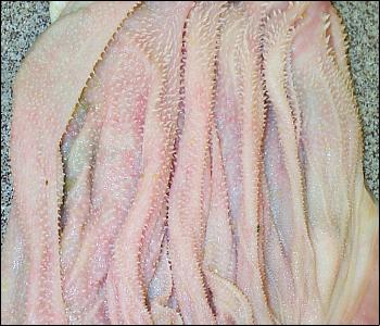 |
The anatomic features described above are exemplified by cattle, sheep and goats. Certain other animals are also generally called ruminants, but have slightly different forestomach anatomy. Camelids (camels, llamas, alpacas, vicunas) have a reticulum with areas of gland-like cells, and an omasum that is tubular and almost indistinct. These animals are occasionally referred to as pseudoruminants or as having "three stomachs" rather than four.
A final note on anatomy. Stratified, squamous epithelium such as found in the rumen is not usually considered an absorptive type of epithelium. Ruminal papillae are however very richly vascularized and the abundant volatile fatty acids produced by fermentation are readily absorbed across the epithelium. Venous blood from the forestomachs, as well as the abomasum, carries these absorbed nutrients into the portal vein, and hence, straight to the liver.
Send comments to Richard.Bowen@colostate.edu